
SI-VMA Automatic Focusing Video Microscope
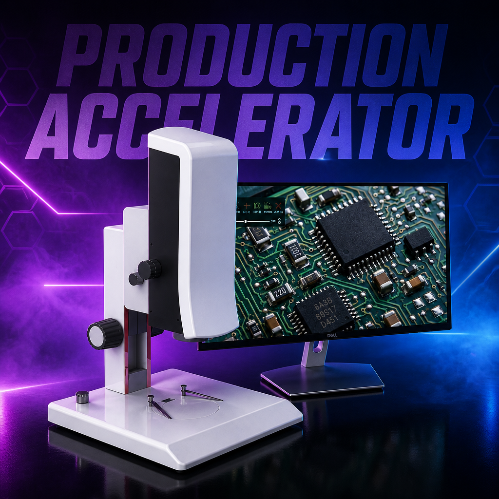
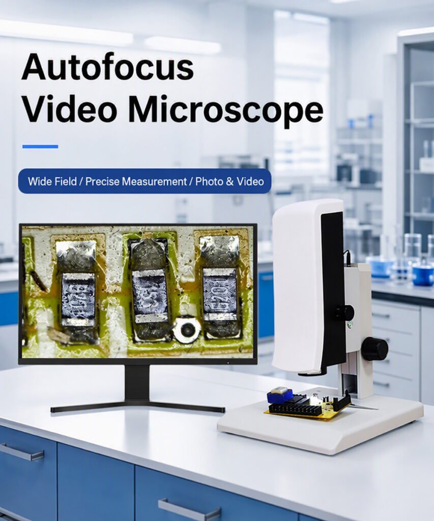
See Every Detail — Without Ever Touching a Focus Knob The SI-VMA Automatic Focusing Video Microscope is a plug-and-use inspection system built for production floors, QC benches and workstations. Move ...
GET QUOTE NOW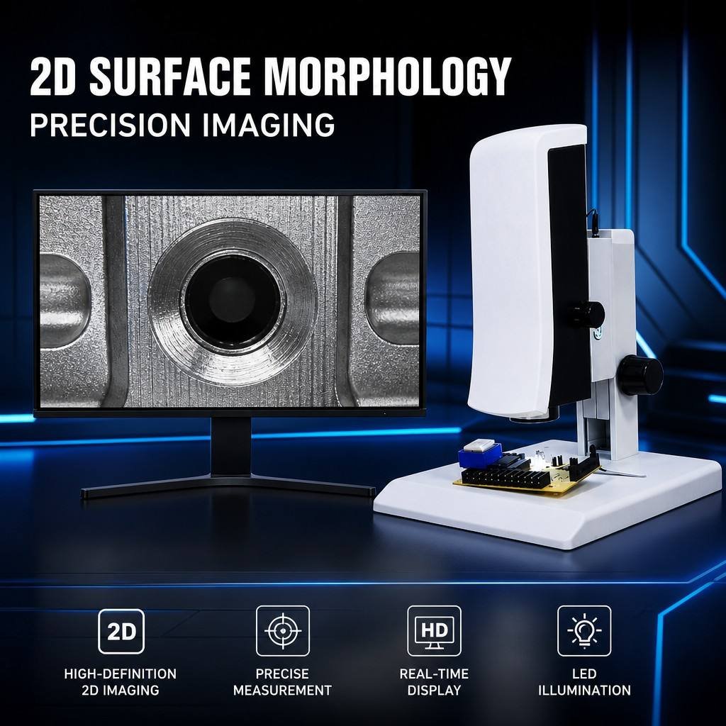
Why Teams Choose the SI-VMA
Why Teams Choose the SI-VMA
⚡ Real-Time Autofocus — Three Focus Modes
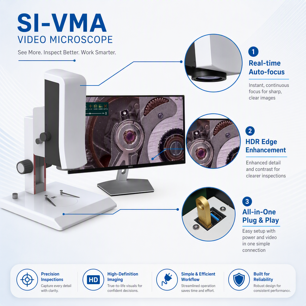
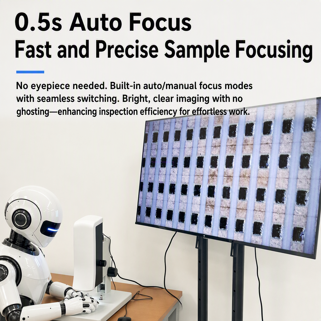
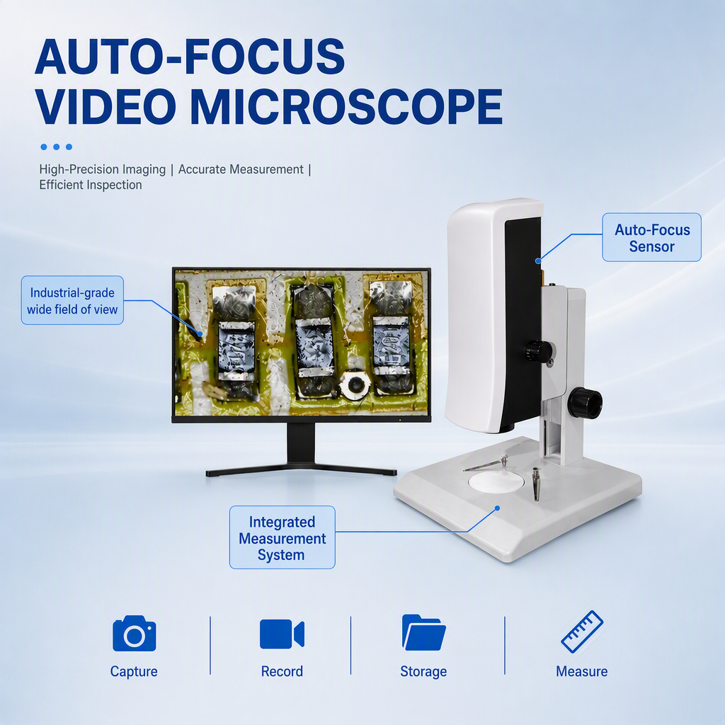
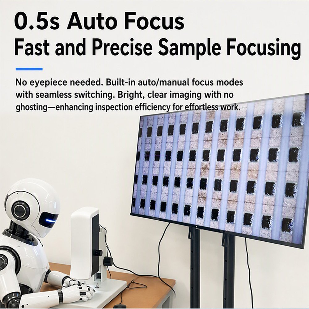
Choose Auto, One-Touch or Manual focus. In Auto mode the system detects a blurred image and refocuses on its own. Enable the on-screen focus frame and simply click anywhere on the image to focus on that point. In Manual mode, drag the focus slider or scroll the mouse wheel for precise control.
🔍 Continuous Optical Zoom + Digital Magnification
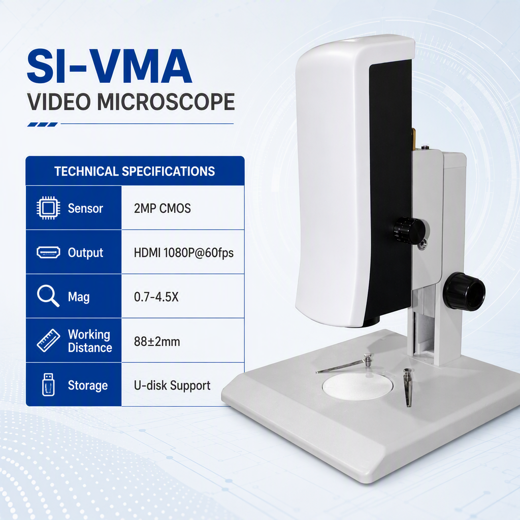
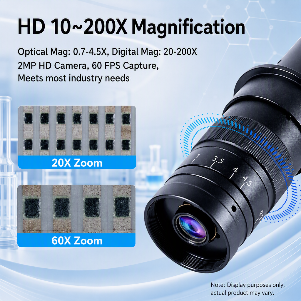
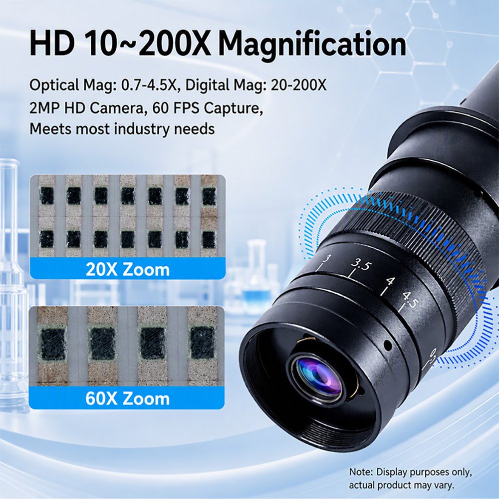
A manual 0.7–4.5X continuous zoom lens gives a system magnification range of 32.9–211.6X. Add 1.0–6.0X electronic magnification on top for detail reproduction up to 180X electronic magnification on integrated circuits. All magnifications are parfocal from the factory, with centre deviation held to < 0.05 mm between magnification steps.
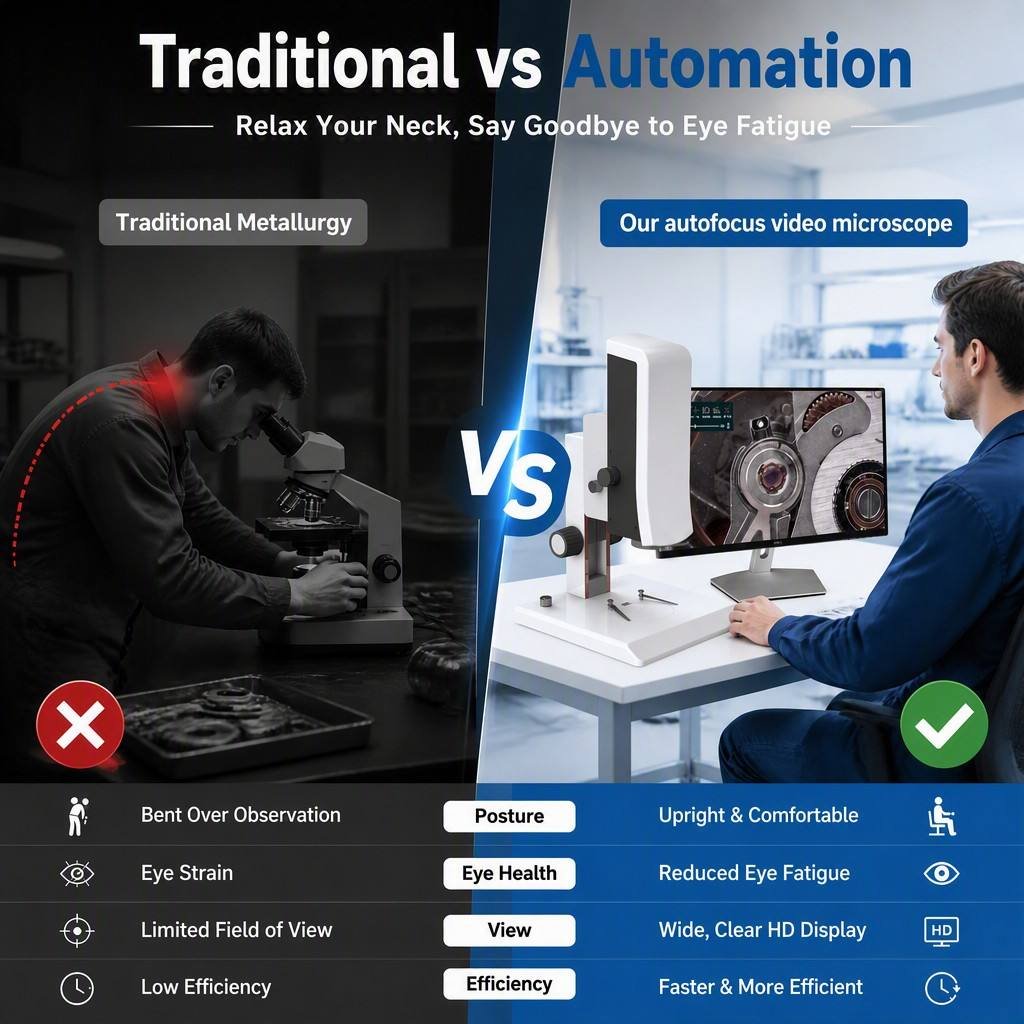
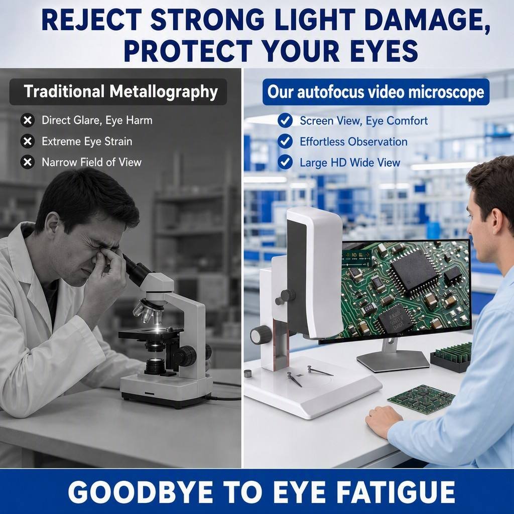
🖥️ Large-Screen HDMI Viewing — No Eyepiece, No Neck Strain
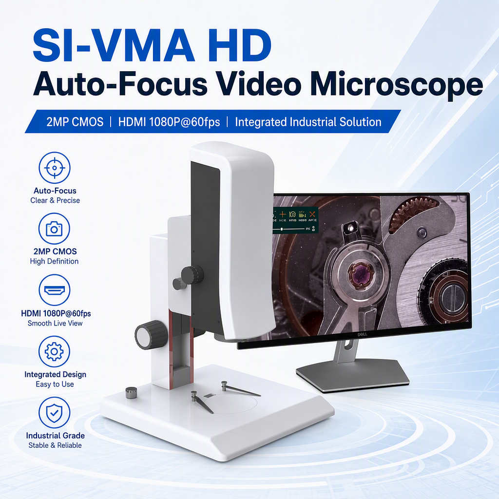
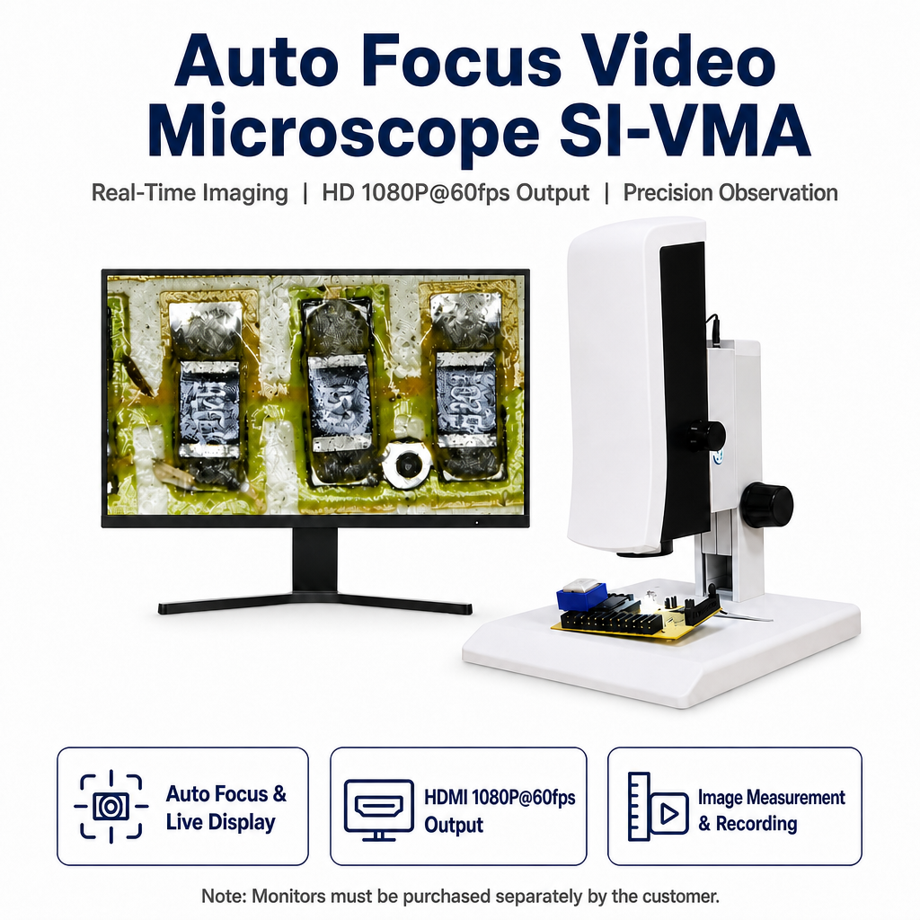
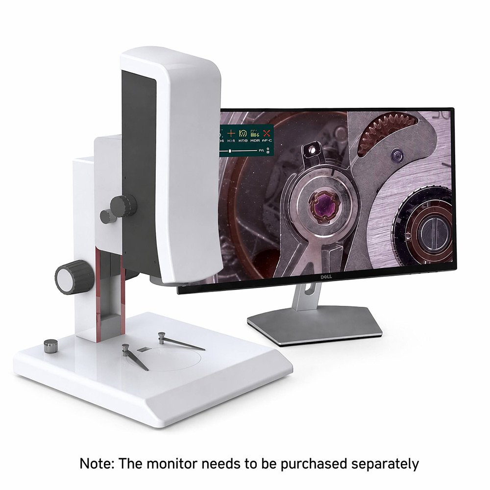
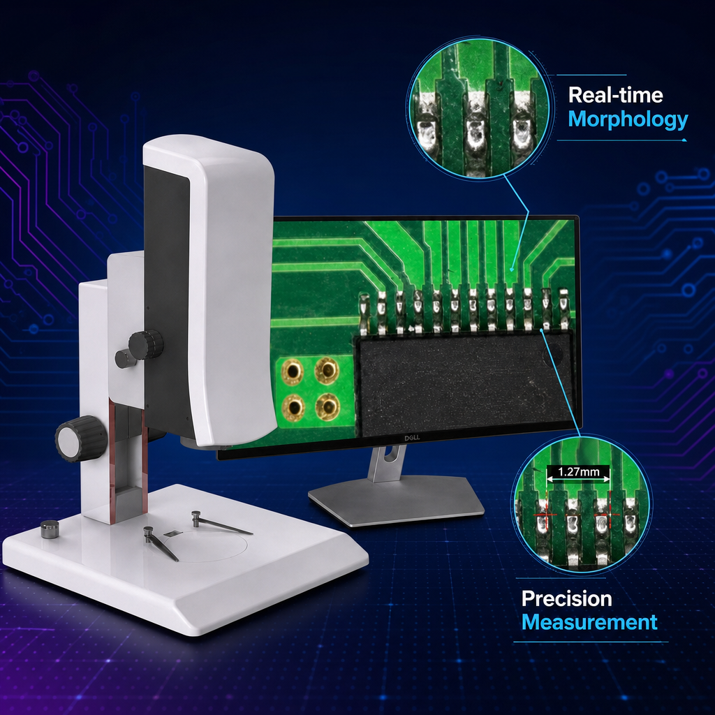
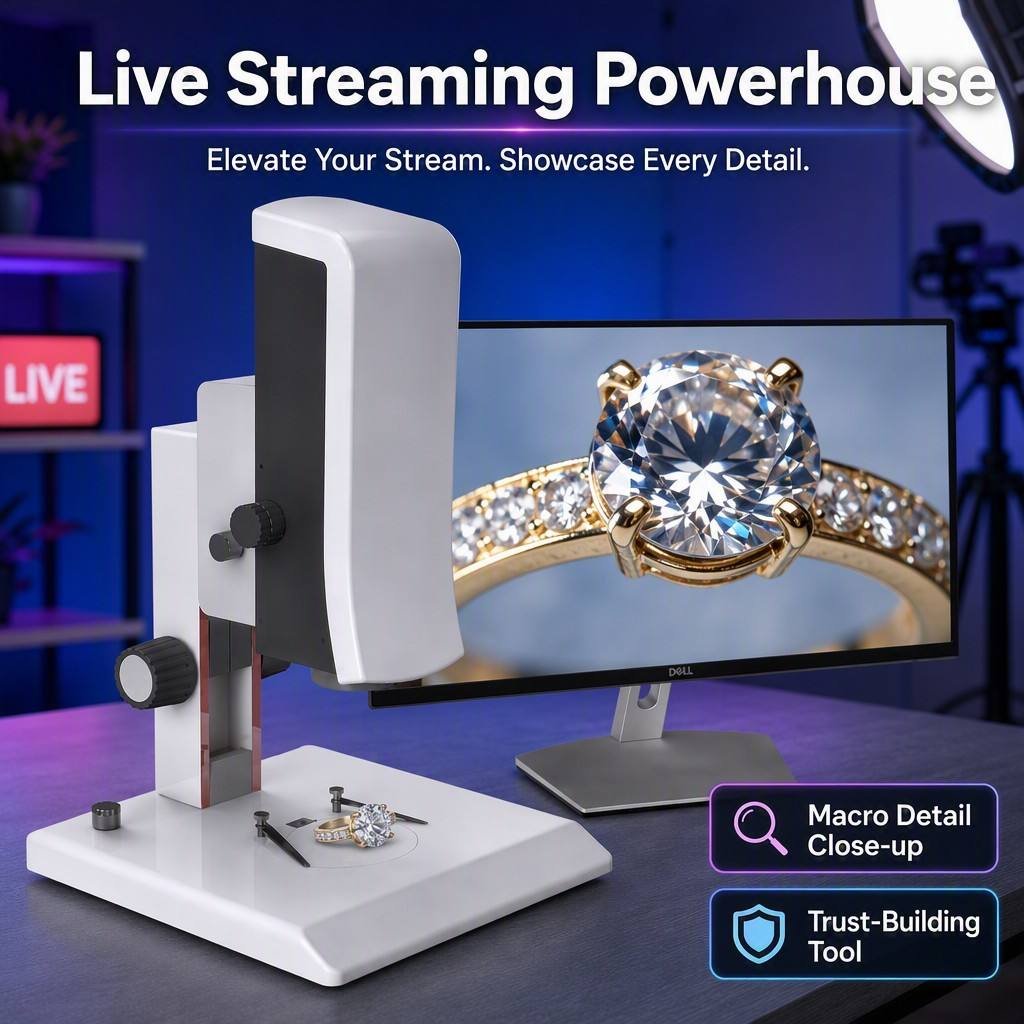
The SI-VMA outputs 1080P over HDMI directly to any monitor. Operators inspect on a large screen instead of hunching over an eyepiece — better for long shifts, better for training, and better when several people need to review the same defect at once. Note: the monitor is not included and must be purchased separately.
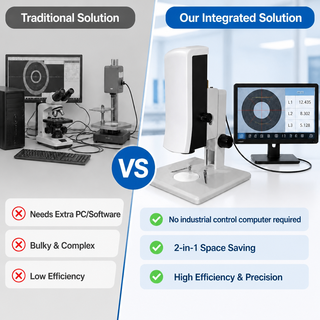
🎯 Built-In Measurement Suite — No PC Software Needed
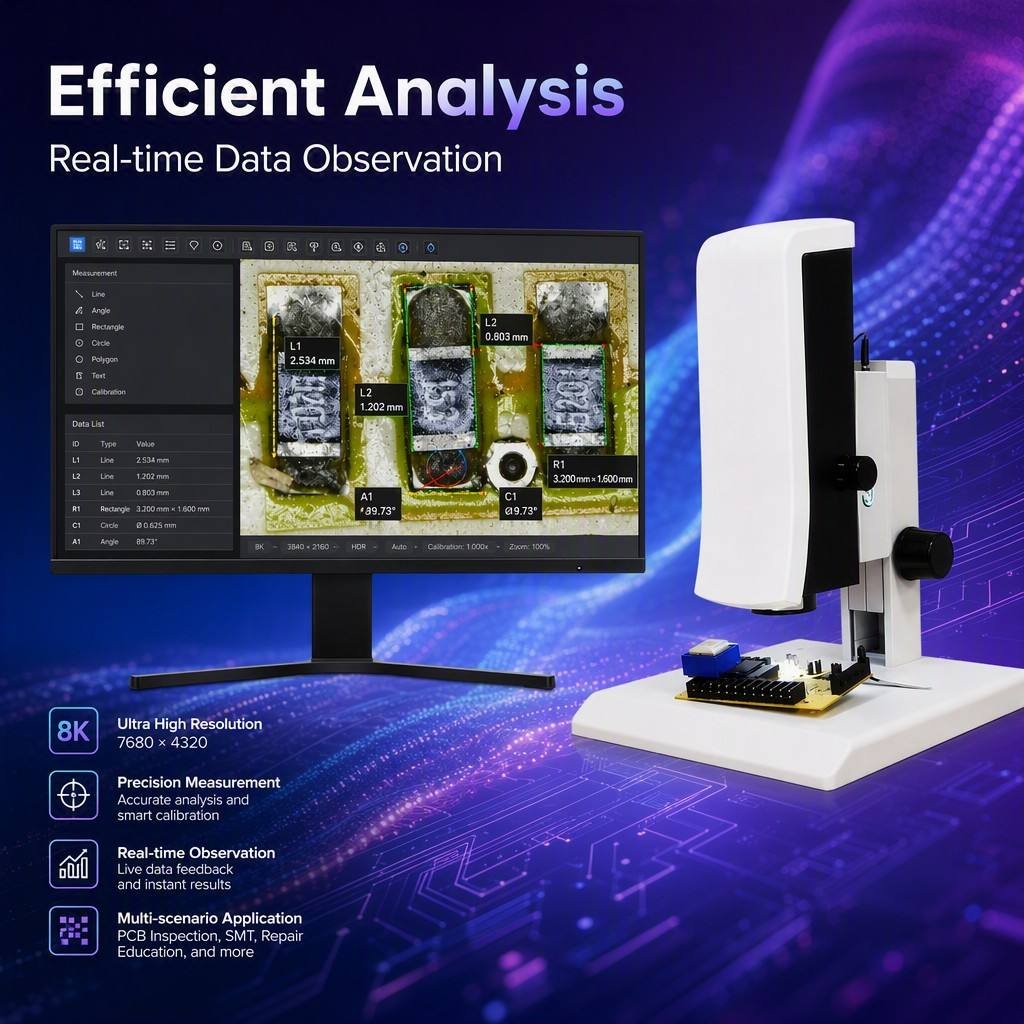
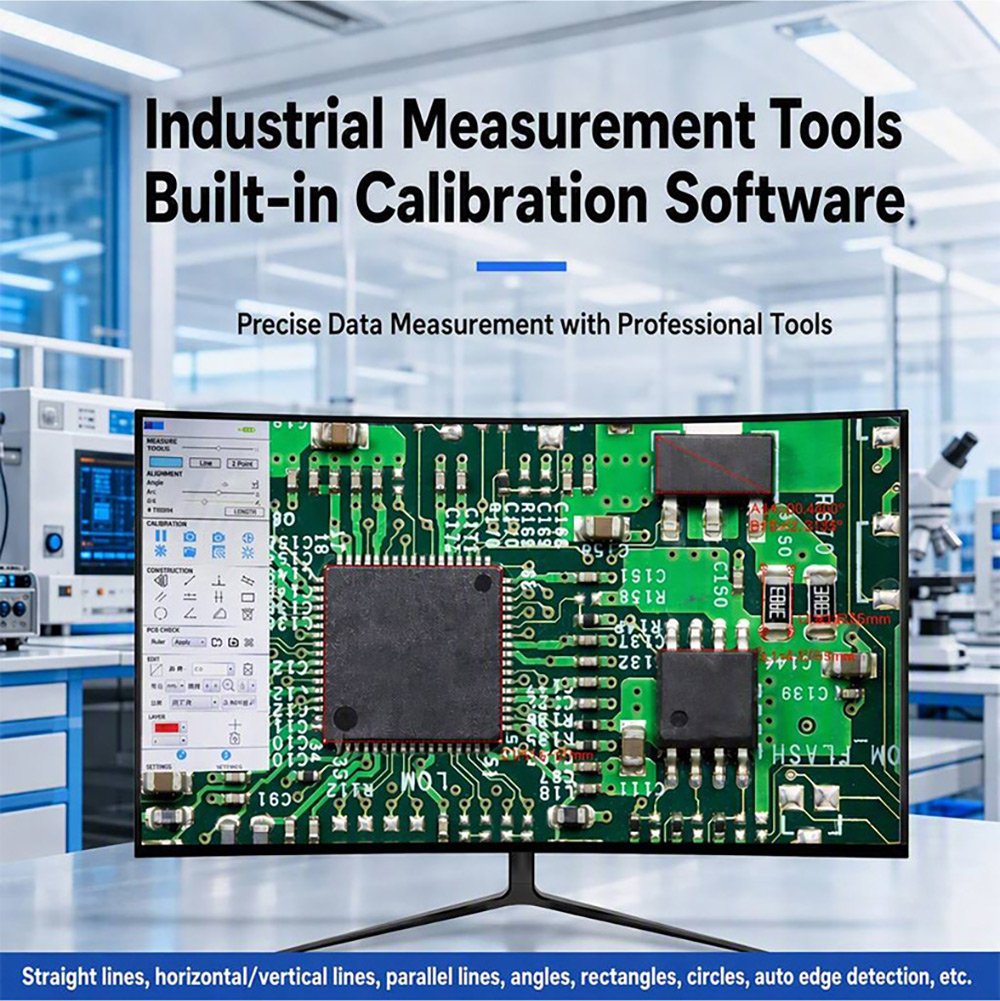
Measure directly on the live image with a full toolset: line · horizontal line · vertical line · rectangle · parallel lines · perpendicular line · angle · polygon · arc · circle · concentric circles · double circle · line-to-circle · perpendicular-to-circle · annotation · scale bar.
Edge detection automatically snaps line, horizontal and vertical measurements to the true edge
Calibration supports linear, H-type and circle calibration with user-defined units
Custom templates — save a repeated measurement layout once and recall it for every part
Export measurement data to a .CSV file on the USB drive with a filename you choose
Barcode workflow: connect a scan gun and the system auto-creates a folder named after the scanned barcode and files all captures inside it
🌈 HDR & Edge Enhancement for Difficult Surfaces
The wide dynamic range (HDR) mode reveals detail in bright and dark regions simultaneously — ideal for shiny solder joints, black plastics and mixed-reflectivity assemblies. Edge enhancement sharpens boundary definition so defects stand out. Note: frame rate drops to 30 fps when wide dynamic range is enabled.
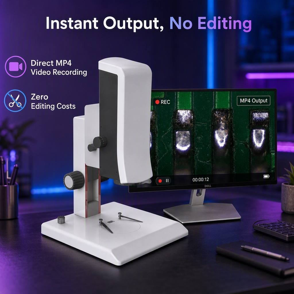
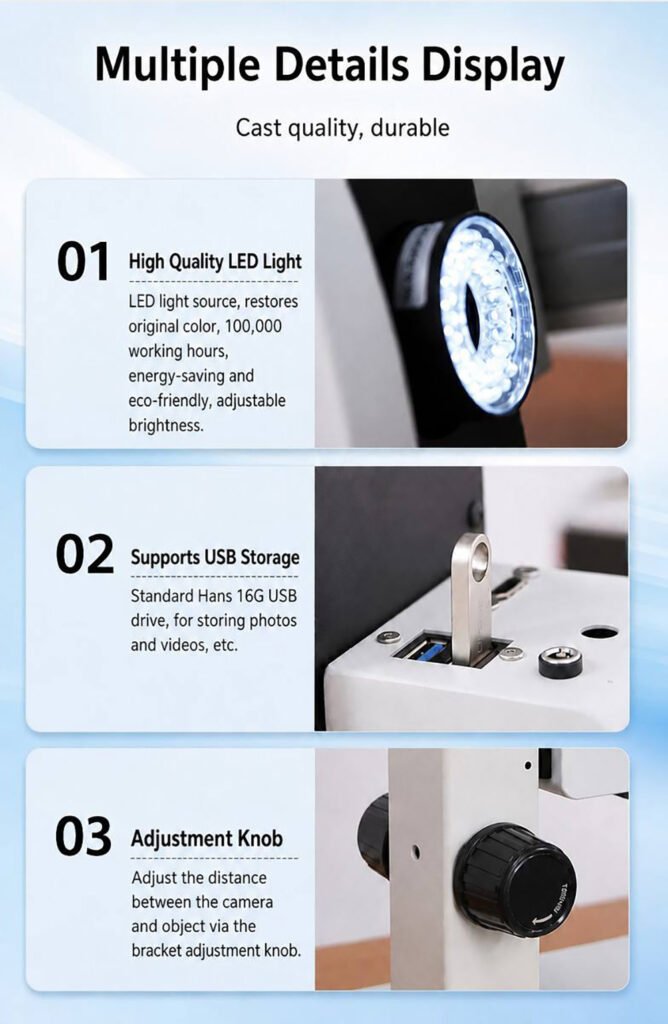
💾 Direct USB Storage — No Computer in the Loop
Insert a USB flash drive and capture immediately:
Photos in .JPG or .BMP, auto-named by timestamp or a custom name
Video in .MP4, up to approximately 2 hours / 4 GB per recording
Measurement data in .CSV
Optional: burn measurement overlays into the saved image (Save Primitives = Yes)
🖱️ Mouse-Driven Interface — Nothing to Install
Operate everything with a standard USB mouse through an on-screen menu. Adjust exposure (auto/manual gain and shutter), white balance (auto/manual RGB), contrast (0–100), sharpness (0–15), saturation (0–255) and gamma (0–3). Additional tools include image freeze, live-vs-saved image comparison (BMP), crosshair and crosshair-scale overlays with adjustable colour, invert / flip, anti-flicker, monochrome mode and factory reset.
💡 Adjustable LED Ring Light
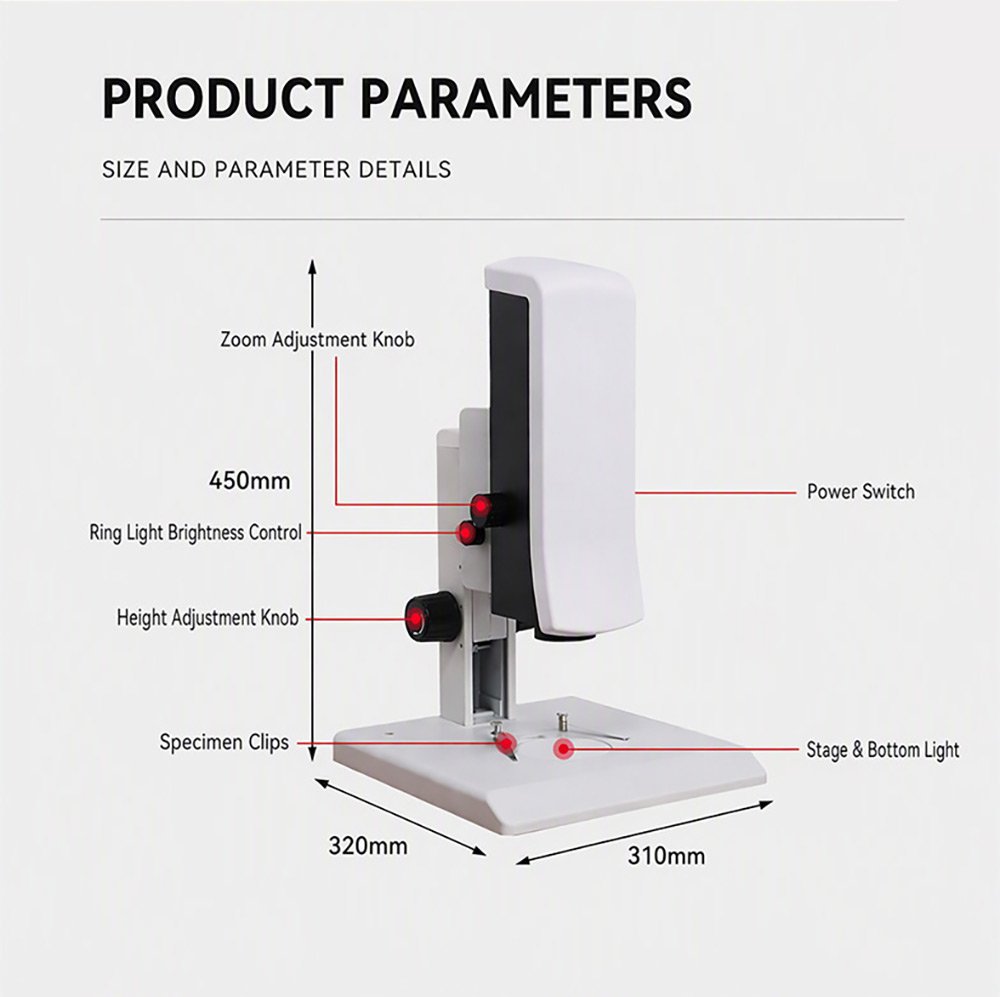
An integrated ring surface light with a brightness control knob provides even illumination. Multiple light-source matching modes are available to suit different sample characteristics.
🔧 Interchangeable Objectives
Independently developed lens mounting supports objective lens replacement, so one base system adapts to different working environments and inspection tasks.
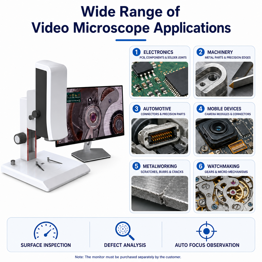
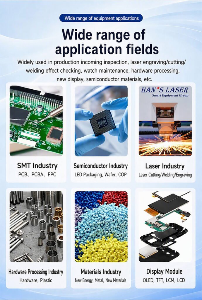
Where the Autofocus Video Microscope Is Used
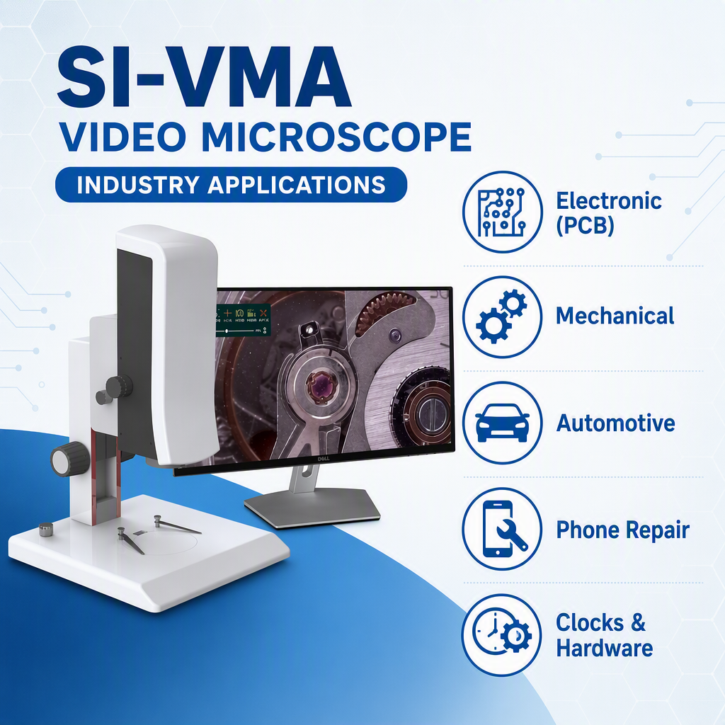
Industry Typical Inspection Tasks
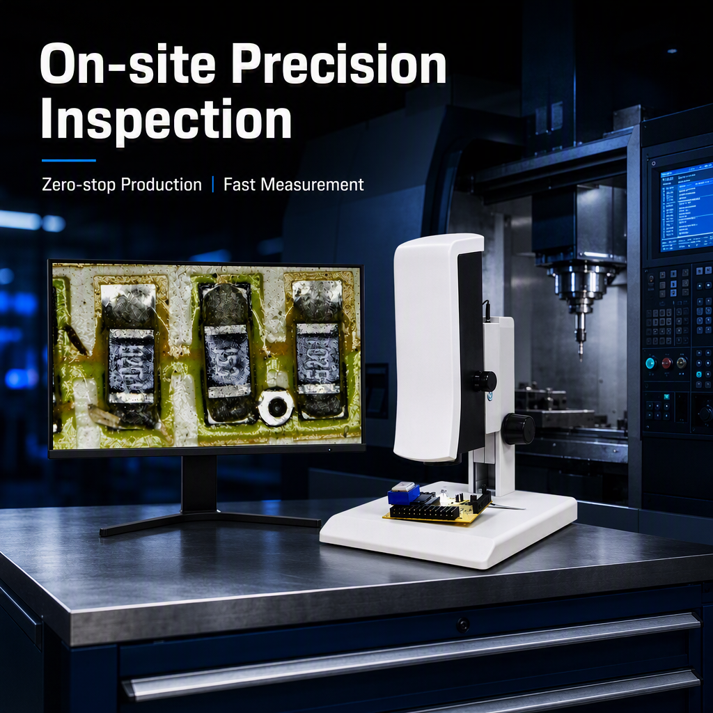
SMT / Electronics PCB, PCBA, FPC — solder joints, components, traces
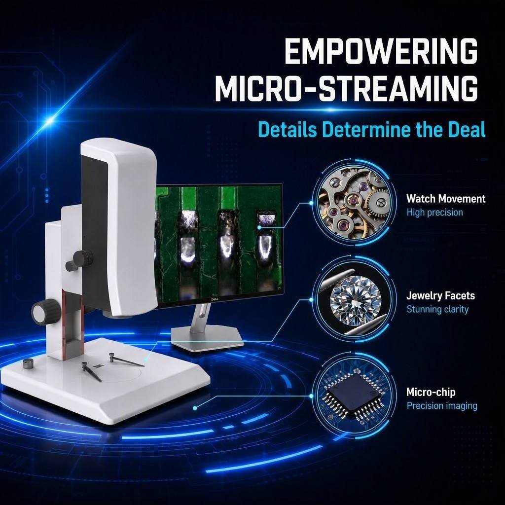
Semiconductor LED packaging, wafer, COP
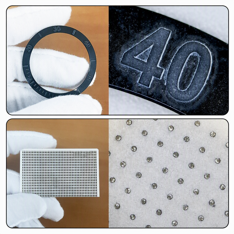
Laser Processing Laser cutting, welding and engraving effect verification
Hardware Processing Metal and plastic parts, precision edges, burrs, scratches, cracks
Materials New energy materials, metals, new material research
Display Modules OLED, TFT, LCM, LCD
Mobile Devices Camera modules, connectors, precision parts
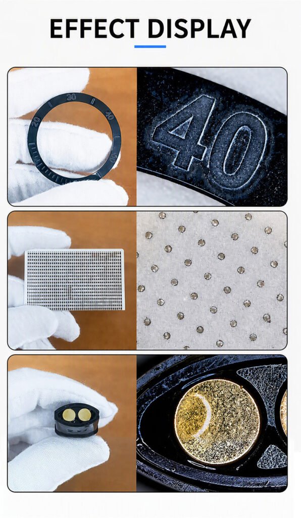
Watchmaking Gears, micro-mechanisms, bezel engraving
Automotive Connectors and precision components
General QC Incoming inspection, in-process inspection, surface inspection, defect analysis
OEM / ODM — Custom Solutions
We support end-to-end customisation through a six-stage workflow:
1.Inquiry & Brief — discuss application, measurement needs and OEM/ODM goals
2.Technical Proposal — confirm magnification, imaging and measurement requirements
3.Design & Sample — develop the customised design and verify sample performance
4.Production — assemble microscope systems under controlled manufacturing
5.Quality Testing — autofocus test, image quality, measurement accuracy, system function, final inspection
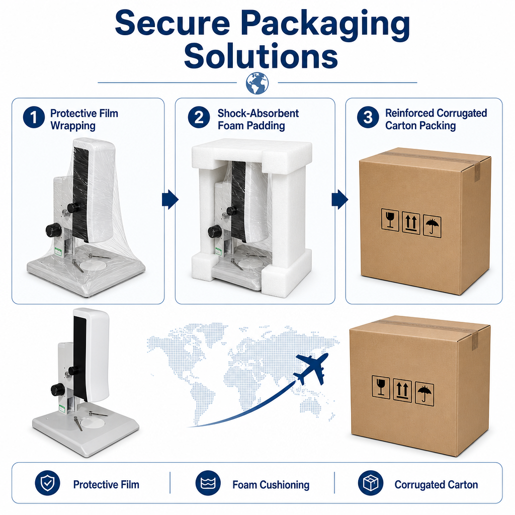
6.Delivery & Support — safe packing plus technical support worldwide
Care & Maintenance
Keep the instrument out of direct sunlight; store in a dry, clean, low-vibration environment
Handle gently — this is a precision instrument; avoid impact during transport
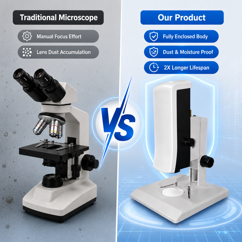
Keep all lens surfaces free of dust and fingerprints
Remove dust with a blower or gentle cotton wipe; remove oil or fingerprints with cotton lightly dampened in a 3:7 ethanol–ether mixture
Clean non-optical surfaces with neutral detergent only — never organic solvents, especially on plastics
Do not disassemble the microscope yourself
Cover with the dust cover when not in use and store away from humidity
Never rotate the left and right zoom hand wheels in opposite directions — this will cause a fault
Periodic inspection is recommended to maintain performance
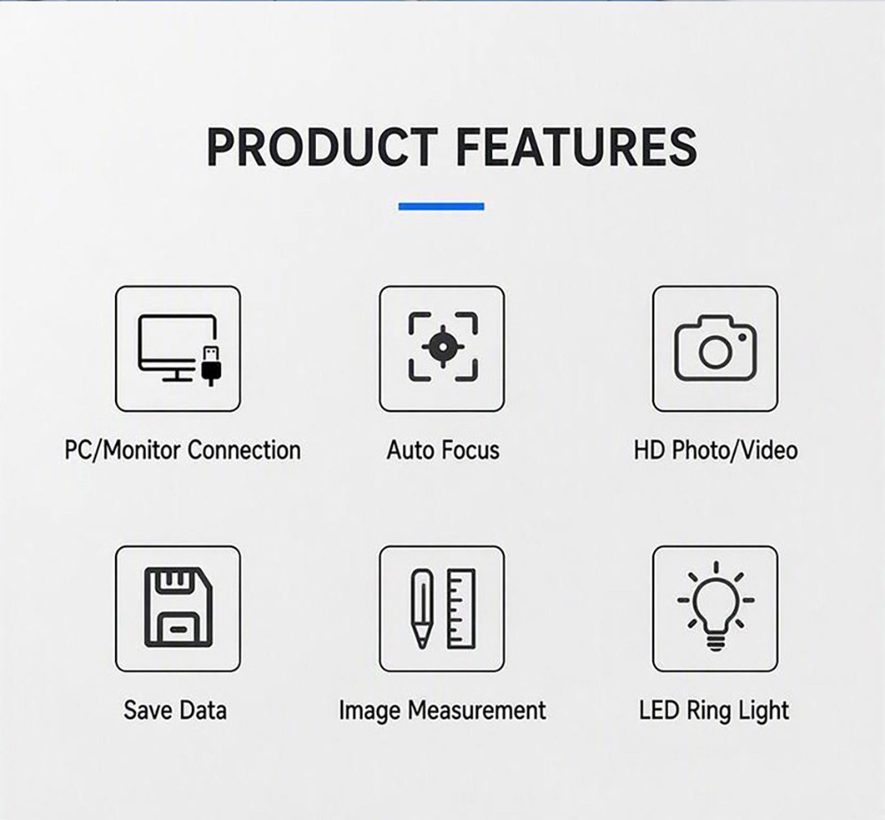
Key Features
Real-Time Autofocus — Three Focus Modes
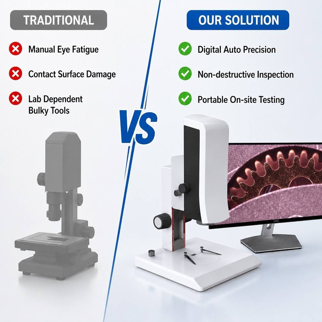
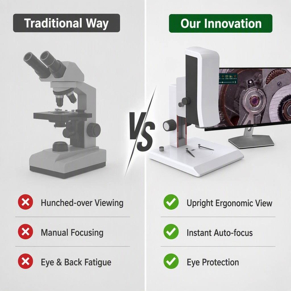
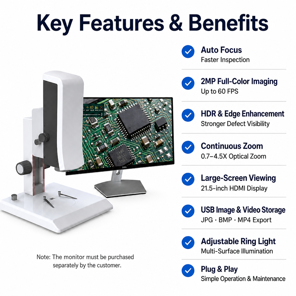
Choose Auto, One-Touch or Manual focus. In Auto mode the system detects a blurred image and refocuses on its own. Enable the on-screen focus frame and simply click anywhere on the image to focus on that point. In Manual mode, drag the focus slider or scroll the mouse wheel for precise control.
Continuous Optical Zoom + Digital Magnification
A manual 0.7–4.5X continuous zoom lens gives a system magnification range of 32.9–211.6X. Add 1.0–6.0X electronic magnification on top for detail reproduction up to 180X electronic magnification on integrated circuits. All magnifications are parfocal from the factory, with centre deviation held to < 0.05 mm between magnification steps.
Large-Screen HDMI Viewing — No Eyepiece, No Neck Strain
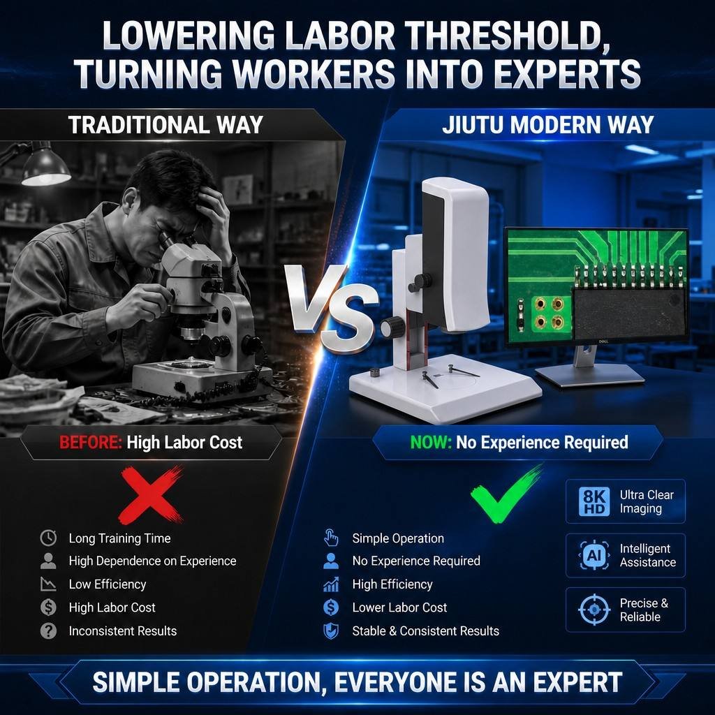
The SI-VMA outputs 1080P over HDMI directly to any monitor. Operators inspect on a large screen instead of hunching over an eyepiece — better for long shifts, better for training, and better when several people need to review the same defect at once. Note: the monitor is not included and must be purchased separately.
Built-In Measurement Suite — No PC Software Needed
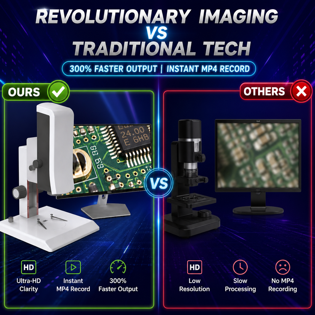
Measure directly on the live image with a full toolset: line · horizontal line · vertical line · rectangle · parallel lines · perpendicular line · angle · polygon · arc · circle · concentric circles · double circle · line-to-circle · perpendicular-to-circle · annotation · scale bar. Edge detection automatically snaps line, horizontal and vertical measurements to the true edge. Calibration supports linear, H-type and circle calibration with user-defined units. Custom templates — save a repeated measurement layout once and recall it for every part. Export measurement data to a .CSV file on the USB drive with a filename you choose. Barcode workflow: connect a scan gun and the system auto-creates a folder named after the scanned barcode and files all captures inside it.
HDR & Edge Enhancement for Difficult Surfaces
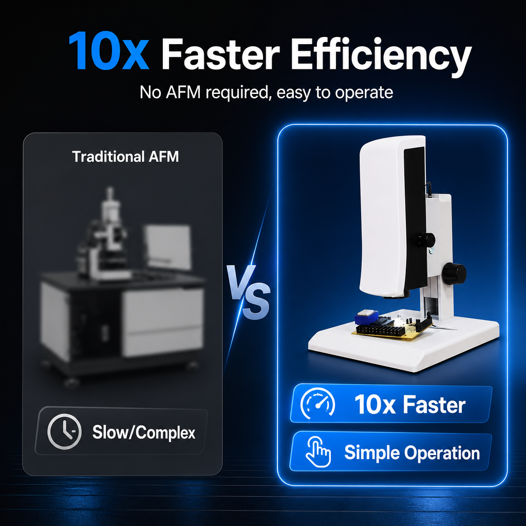
The wide dynamic range (HDR) mode reveals detail in bright and dark regions simultaneously — ideal for shiny solder joints, black plastics and mixed-reflectivity assemblies. Edge enhancement sharpens boundary definition so defects stand out. Note: frame rate drops to 30 fps when wide dynamic range is enabled.
Direct USB Storage — No Computer in the Loop
Insert a USB flash drive and capture immediately: Photos in .JPG or .BMP, auto-named by timestamp or a custom name. Video in .MP4, up to approximately 2 hours / 4 GB per recording. Measurement data in .CSV. Optional: burn measurement overlays into the saved image (Save Primitives = Yes).
Mouse-Driven Interface — Nothing to Install
Operate everything with a standard USB mouse through an on-screen menu. Adjust exposure (auto/manual gain and shutter), white balance (auto/manual RGB), contrast (0–100), sharpness (0–15), saturation (0–255) and gamma (0–3). Additional tools include image freeze, live-vs-saved image comparison (BMP), crosshair and crosshair-scale overlays with adjustable colour, invert / flip, anti-flicker, monochrome mode and factory reset.
Adjustable LED Ring Light
An integrated ring surface light with a brightness control knob provides even illumination. Multiple light-source matching modes are available to suit different sample characteristics.
Interchangeable Objectives
Independently developed lens mounting supports objective lens replacement, so one base system adapts to different working environments and inspection tasks.
Why Choose Us
OEM / ODM — Custom Solutions
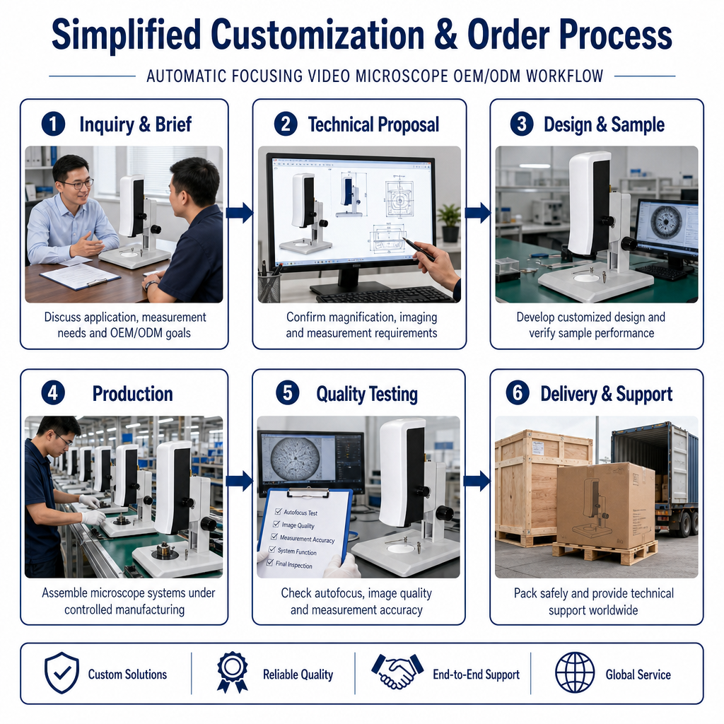
We support end-to-end customisation through a six-stage workflow:
1.Inquiry & Brief — discuss application, measurement needs and OEM/ODM goals 2.Technical Proposal — confirm magnification, imaging and measurement requirements 3.Design & Sample — develop the customised design and verify sample performance 4.Production — assemble microscope systems under controlled manufacturing 5.Quality Testing — autofocus test, image quality, measurement accuracy, system function, final inspection 6.Delivery & Support — safe packing plus technical support worldwide
Product Documents
Shengmei Instrument - Autofocus Video Microscope - Product Description (EN)
Shengmei Instruments - Autofocus Video Microscope - Operating Instructions V2.0 (HDMI)
Key Features
Product Details

FAQ
Q1. What magnification does the SI-VMA reach?
Q2. Is a monitor included?
Q3. Do I need a computer or driver software?
Q4. Is there an eyepiece?
Q5. Can the objective lens be changed?
Q6. How does the autofocus actually work?
Q7. Can I measure on screen, and what shapes are supported?
Q8. Where are files saved?
Q9. Do you support OEM / ODM?
Q10. How is the microscope packed for shipping?


Interested in This Product?
Tell us your project details and our team will prepare a customized quotation.
GET QUOTE NOW